Diagnosis of urethral strictures requires a careful evaluation of the patient’s clinical history, a physical examination of the patient, as well as various clinical investigations.
Uro-flowmetry
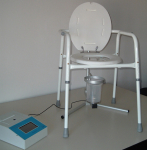
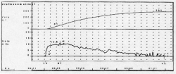
| Uro-flowmetry: A simple investigation that can be performed in the urological out-patient office. The patient urinates normally into a special container connected to a computer, that registers the volume and velocity of the urine stream. To perform this test, no specific preparation of the patient is required, except for a full bladder in order to urinate. | ||
Ultrasonography
Ultrasonography: The exam is performed after the uroflowmetry and helps evaluate whether the patient succeeded in completely emptying the bladder by urinating. The presence of urine in the bladder after micturition can be related to urethral stricture.
Urethrography
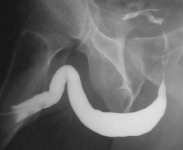
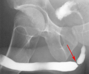
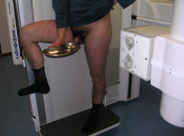
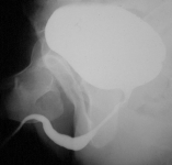
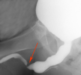
Urethrography: This radiological examination of the urethra is performed with the use of contrast medium. Retrograde and voiding urethrography is the most basic exam for studying the urethra. Non- urethral surgery can be planned without having performed this exam. The exam is composed of two phases:
|
||
|
||
Urethral Ultrasonography
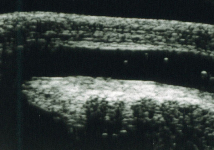
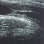
| Urethral ultrasonography: This exam is performed using an auxiliary ultrasonic probe that is placed on the penis or perineum. It is a simple and entirely painless exam, which does not require any specific patient preparation. | |
Urethroscopy
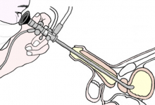
| Urethroscopy: This procedure is performed in an operating room with the patient under anesthesia to allow for an easy and painless exam. The examination consists in introducing a metal instrument into the urethra in order to visualize the urethral canal from the inside. The instrument, which can come in different sizes, is inserted into the external urethral meatus and is carefully moved through the urethra until it reaches the bladder. | ||
In some patients, further examinations may be required such as CT, NMR, Intravenous Pyelogram (IVP), and Urodynamics test.

