Dorsal oral mucosal onlay graft urethroplasty (Asopa’s technique)

  • Introduction
  • Lectures
  • Articles
  • F.A.Q.
  • Results
  • Surgical Techniques
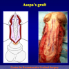
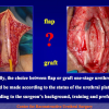
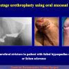
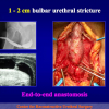
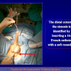
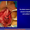
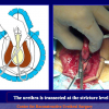
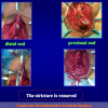
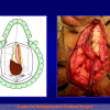
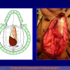
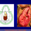
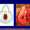
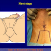
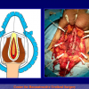
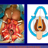
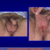
The surgical technique of “Urethroplasty with skin or oral mucosal graft” is made by opening the urethra at the level of the stricture site and widening the urethral lumen by applying an oral graft.

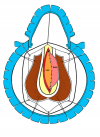
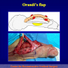
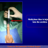
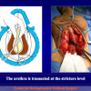
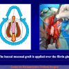
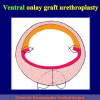
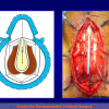
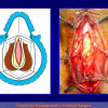
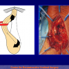
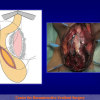
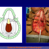
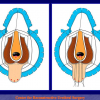
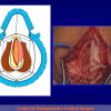
Figure 1

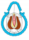
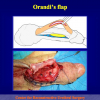
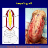
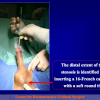
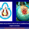
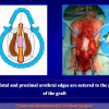
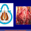
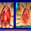
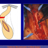
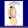
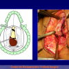
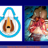
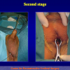
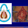
Figure 2

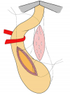
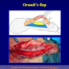
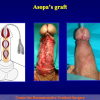
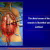
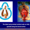
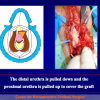
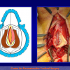
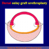
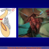
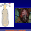
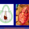
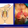
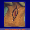
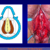
Figure 3

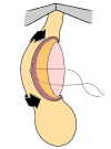
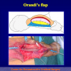
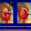
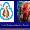
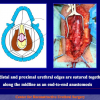
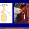
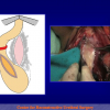
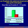
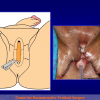
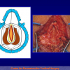
Figure 4

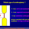
There are four basic types of Urethroplasty with skin or oral mucosal graft:

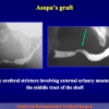
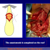
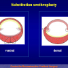
  • Urethroplasty with ventral oral mucosal graft. Using this technique, the oral graft is sutured on the ventral site of the urethra (figures 1,2).
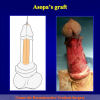
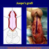
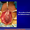
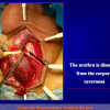
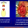
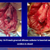
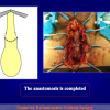
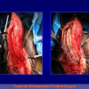
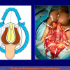
  • Urethroplasty with dorsal skin or oral mucosal graft. Using this technique, the oral graft is sutured on the dorsal site of the urethra (figures 3,4).
  • Urethroplasty with lateral oral mucosal graft. Using this technique, the oral graft is sutured on the lateral site of the urethra.
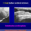
  • Urethroplasty with complete substitution by oral mucosal graft. Using this technique, the urethral mucosa is completely replaced by an oral graft.


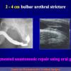
The surgical technique of Urethroplasty with skin or oral mucosal graft is generally suggested in the patients with no traumatic bulbar urethral strictures, more than 2 cm in length.

Article n° 1
Barbagli G, Selli C, di Cello V, Mottola A.
A one-stage dorsal free-graft urethroplasty for bulbar urethral strictures.
Br J Urol. 1996 Dec;78(6):929-32

Objective: To report the use of one-stage dorsal free-graft urethroplasty to reduce the incidence of urethrocele. Patients and Methods: From 1990 to 1994, 20 men (age range 21-86 years) underwent a one-stage dorsal free-graft urethroplasty of bulbar urethral strictures (iatrogenic in 12, traumatic in three, inflammatory in three and unknown in two). All patients except one had been treated previously by optical urethrotomy from one to 14 times.
Results: Temporary fistulae were detected on post-operative urethrography in three patients with particularly long grafts, but they all resolved spontaneously. Within a mean follow-up of 46 months, only one patient had a short recurrent stricture, which was treated successfully by optical urethrotomy. Two patients complained of post-voiding dribbling, but radiographic studies never showed graft weakening and the urinary flow rate was always > 14 mL/s.
Conclusions: Free skin grafts can be applied successfully to the dorsal aspect and by doing so the complications of urethral reconstruction can be reduced.

Article n° 2
Barbagli G, Selli C, Tosto A, Palminteri E.
Dorsal free graft urethroplasty.
J Urol. 1996 Jan;155(1):123-6.

Purpose: Dorsal free graft urethroplasty was performed to reduce the incidence of urethrocele.
Materials and Methods: We treated 12 patients with penile and 13 with bulbous strictures. Of the 13 patients with a bulbous stricture 6 received a dorsally placed tube graft and 7 received a patch graft.
Results: Temporary fistulas were seen on postoperative urethrography in 5 cases but they all resolved spontaneously. At a mean followup of 35.8 months clinical and radiological findings were excellent in 23 cases and good in 2. No signs of graft weakening, such as post-void dribbling or diminished ejaculation, were apparent.
Conclusions: The use of free skin grafts for urethral reconstruction is anatomically healthier in the dorsal than in the ventral position.

Article n° 3
Barbagli G, Palminteri E, Rizzo M.
Dorsal onlay graft urethroplasty using penile skin or buccal mucosa in adult bulbourethral strictures.
J Urol. 1998 Oct;160(4):1307-9.

Purpose: Preputial skin graft is used routinely for urethral reconstruction in patients with stricture disease. Alternative donor sites include extrapenile skin, bladder mucosa and buccal mucosa. Recently buccal mucosa graft has been suggested when local epithelial tissue is not available. We describe our experience with 37 patients undergoing 1-stage correction of bulbar urethral stricture using a penile skin (31) or buccal mucosa (6) graft.
Materials and Methods: In 37 patients with bulbar urethral strictures a nontubularized dorsal onlay graft was used for urethral reconstruction. A preputial skin graft was used in 31 patients and a buccal mucosa graft in 6 with a paucity of local skin. Buccal mucosa graft length ranged from 2.5 to 5 cm. (average 4) and preputial skin graft was 2.5 to 12 cm. long (average 4.7). A dorsal approach to the urethral lumen was used in all patients who underwent onlay graft urethroplasty.
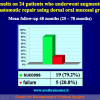
Results: Mean followup was 21.5 months for all 37 patients, 23 months for 31 treated with preputial skin graft and 13.5 months for 6 treated with buccal mucosa graft. The clinical outcomes were considered a failure anytime postoperative instrumentation was needed, including dilatation. In the series 34 cases (92%) were classified as a success and 3 (8%) as failure.
Conclusions: Onlay graft urethroplasty provided excellent results in 92% of adults with bulbourethral stricture. The dorsal approach to the urethra allowed the use of foreskin or buccal mucosa graft for reconstruction of the adequate urethral lumen.

Article n° 4
Palminteri E, Lazzeri M, Guazzoni G, Turini D, Barbagli G.
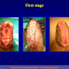
New 2-stage buccal mucosal graft urethroplasty.
J Urol. 2002 Jan;167(1):130-2.

Purpose: Previously buccal mucosal grafts used for repairing adult bulbourethral stricture with the 1-stage dorsal technique has provided a satisfactory outcome in our experience. We present the wider use of buccal mucosal grafts for 2-stage urethroplasty.
Materials and Methods: A total of 24 men 25 to 60 years old (median age 45) with a complex bulbar stricture underwent 2-stage urethroplasty using a buccal mucosal graft to repair the perineostomy. The primary etiology of stricture was traumatic in 4 cases, inflammatory in 16 and unknown in 4. The 2 x 6 cm. graft was harvested from the inner cheek and sutured to the left margin of the urethral mucosal plate with running 6-zero polyglactin suture. Patients were discharged from the hospital within 3 days with a 14Fr silicone urethral catheter in place. Radiological studies and urethroscopy were done 1 year after closure.
Results: A final successful outcome with no recurrent stricture was achieved in 23 of 24 men (92.8%) at a median followup of 18 months (range 13 to 32). In 1 case a urethrocutaneous fistula at the initial radiological assessment closed spontaneously after 14 days of catheterization. No urethral diverticula developed. The mean postoperative peak flow rate is 22 ml. per second (range 18 to 25).
Conclusions: Our new 2-stage buccal mucosal graft urethroplasty may be an excellent technique for complex bulbar urethral stricture disease. Our suggestions may increase usefulness of the 2-stage technique for repairing complex strictures due to the avoidance of classic complications.

Article n° 5
Barbagli G, Palminteri E, Balò S, Picinotti A, Lazzeri M,
Dorsal onlay graft urethroplasty. Current technique step-by-step.
Contemporary Urology, 2002: 14(5), 18-32

The ideal surgical procedure for long bulbourethral strictures would be simple, reliable, effective over the long term, and reproducible in the hands of any surgeon. The Barbagli procedure — based on sound atomic, historic, and surgical principles — fulfils these criteria.

Article n° 6
Barbagli G, Palminteri E, Lazzeri M, Bracka A.
Penile and bulbar urethroplasty using dorsal onlay techniques.
Atlas Urol Clin., 2003: 11, 29-41

The surgical treatment of adult anterior urethral strictures has been a constantly evolving process. Recently, considerable changes have been introduced: the wider use of the buccal mucosa graft and the use of the dorsal onlay approach, also named the Barbagli procedure. Moreover, in penile urethra, the dorsal placement of the graft is now combined by the incision of the urethral plate, as suggested by Snodgrass for childhood hypospadias surgery and its augmentation, as suggested by Bracka, Asopa and Hayes.
Three different techniques are presented:
Penile one-stage urethroplasty with urethral plate incision and augmentation using a dorsal buccal mucosa graft;
Bulbar one-stage urethroplasty with urethral plate augmentation using a dorsal buccal mucosa graft;
Bulbar one-stage urethroplasty with urethral place replacement using a dorsal buccal mucosa graft.

Article n° 7
Barbagli G, Palminteri E, Guazzoni G, Montorsi F, Turini D, Lazzeri M.
Interim outcomes of dorsal skin graft bulbar urethroplasty.
JUrol. Volume 172, Issue 4, Pages 1365-1367 (October 2004)

Purpose: We update our interim results of bulbar urethroplasty using a skin graft placed on the dorsal urethral surface.
Materials and Methods: A total of 45 patients with an average age of 45 years underwent dorsal onlay skin graft urethroplasty between January 1994 and December 2000. Of the patients 23 had undergone an average of 2.6 prior endoscopic procedures (range 1 to 14). Preoperative evaluation include clinical history, physical examination, retrograde and voiding urethrography, and ultrasonography. In all patients the bulbar urethra was opened along its dorsal surface, the graft was sutured, splayed and quilted to the corpora cavernosa, and the urethra was rotated to cover the graft. In all patients was used penile skin as substitution material. Mean graft length was 4.7 cm (range 2.5 to 11). Three weeks after surgery voiding cystourethrography was performed.
Results: Average followup was 71 months (range 41 to 110). Clinical outcome was considered a failure when postoperative instrumentation was needed, including dilation. Of 45 cases 33 (73%) were classified as successful and 12 (27%) were failures. The 12 failures were treated with internal urethrotomy (1), end-to-end-anastomosis (1), skin graft urethroplasty (2) and 2-stage urethroplasty (6). Six of the 12 initial failures had a satisfactory final outcome. The remaining 6 patients refused further surgical procedures and received a definitive perineal urethrostomy.
Conclusions: Penile skin grafts used as a dorsal onlay for bulbar urethral reconstruction in a homogeneous series of patients showed a tendency to deteriorate with time. Longer followup is required to compare penile skin with buccal mucosa as substitute materials for bulbar urethral reconstruction.

Article n° 8
Barbagli G, Palminteri E, Guazzoni G, Montorsi F, Turini D, Lazzeri M.
Bulbar urethroplasty using buccal mucosa grafts placed on the ventral, dorsal or lateral surface of the urethra: are results affected by the surgical technique?
J Urol. 2005 Sep;174(3):955-7;

Purpose: The use of buccal mucosa graft onlay urethroplasty represents the most widespread method of bulbar urethral stricture repair. The graft may be placed on the ventral or dorsal urethral surface according to surgeon experience and preference. We investigated whether the results are affected by the surgical technique by comparing the outcome of 3 types of bulbar urethroplasty using buccal mucosa graft.
Materials and Methods: We repaired 50 bulbar urethral strictures with buccal mucosa grafts from 1997 to 2002. Mean patient age was 42 years. The etiology of stricture was ischemia in 12 cases, trauma in 6, instrumentation in 4 and unknown in 28. Patients with lichen sclerosus, failed hypospadias or urethroplasty and stricture extending into the penile urethra were not included. A total of 47 patients (94%) had undergone previous urethrotomy or dilation. The buccal mucosa graft was always harvested from the cheek using a 2 team approach. Mean graft length was 4.2 cm. The graft was placed on the ventral, dorsal and lateral bulbar urethral surface in 17, 27 and 6 cases, respectively. Clinical outcome was considered a success or failure at the time that any postoperative procedure was needed, including dilation. Mean followup was 42 months (range 12 to 76).
Results: Of 50 cases 42 (84%) were successful and 8 (16%) failed. The 17 ventral grafts provided success in 14 cases (83%) and failure in 3 (17%). The 27 dorsal grafts provided success in 23 cases (85%) and failure in 4 (15%). The 6 lateral grafts provided success in 5 cases (83%) and failure in 1 (17%). No surgical complications were observed. Failures involved the anastomotic site (distal in 2 and proximal in 3) and the whole grafted area in 3 cases. They were treated with urethrotomy in 5 cases and 2-stage urethroplasty in 3. Conclusions: In our experience the placement of buccal mucosa grafts into the ventral, dorsal or lateral surface of the bulbar urethra showed the same success rates (83% to 85%) and the outcome was not affected by the surgical technique. Moreover, stricture recurrence was uniformly distributed in all patients.

Article n° 9
Barbagli G, De Stefani S, Sighinolfi MC, Annino F, Micali S, Bianchi G.
Bulbar urethroplasty with dorsal onlay buccal mucosal graft and fibrin glue.
Eur Urol. 2006 Sep;50(3):467-74

Objectives: We describe a new surgical technique with the use of fibrin glue for bulbar urethra reconstruction using a dorsal buccal mucosal onlay graft.
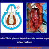
Methods: Six patients with a mean age of 43 yr underwent bulbar urethroplasty with dorsal onlay buccal mucosal graft and fibrin glue. The urethra was mobilised from the corpora cavernosa and opened along its dorsal surface. The buccal mucosal graft was applied on the corpora cavernosa using 2ml of fibrin glue. Two interrupted polyglactin 5-0 sutures were used to fix the apices of the graft to the underlying albuginea of the corpora cavernosa. The urethra was rotated back to cover the graft and an adjunctive fibrin glue was injected over the urethra.
Results: The mean operative time was 100min (range, 90-120min). No intraoperative or postoperative complications were observed. Voiding cystourethrography was performed when the catheter was removed 2 wk after surgery. Urine culture, uroflowmetry, and urethrography were repeated after 6 and 12 mo and annually thereafter. Mean follow-up was 16 mo (range, 12-24 mo). No restrictures at the anastomotic sites were demonstrated in any of the patients 6 and 12 mo after surgery.
Conclusions: The use of fibrin glue represents a slight but significant step toward perfecting the surgical technique of bulbar urethral reconstruction.

Article n° 10
Barbagli G, De Stefani S, Sighinolfi MC, Pollastri CA, Annino F, Micali S, Bianchi G.
Experience with fibrin glue in bulbar urethral reconstruction using dorsal buccal mucosa graft.
Urology. 2006 Apr;67(4):830-2.

Buccal mucosa dorsal onlay graft urethroplasty represents a widespread method for bulbar urethral stricture repair. We describe a modified procedure with the use of fibrin glue applied on the receiving bed before graft location.

Article n° 11
Guido Barbagli, Giuseppe Morgia, Massimo Lazzeri c
Dorsal Onlay Skin Graft Bulbar Urethroplasty: Long-Term Follow-Up
Eur. Urol. 2008,53:628 — 634

Objectives: To report retrospectively long-term follow-up in a homogeneous group of patients who underwent dorsal onlay skin graft bulbar urethroplasty and to investigate which factors might influence longterm outcome.
Methods: Thirty-eight patients, with an average age of 43 yr, underwent dorsal onlay skin graft (#12 ventral penile skin and #26 preputial mucosa) bulbar urethroplasty from 1994 to 2000. Of 38 patients, 23 (60.5%) had undergone prior endoscopic procedures. Preoperative evaluation included clinical history, physical examination, retrograde and voiding urethrography, and urethral sonography. Three weeks after surgery, voiding cystourethrography was performed. Patients were followed-up with a clinical evaluation and specific diagnostic tests every 4 mo in the first year and every 12 mo thereafter. Clinical outcome was considered a failure when postoperative instrumentation, including dilation, was needed.
Results: Average follow-up was 111 mo (range, 80–149). Of 38 cases, 25 (65.8%) were successful and 13 (34.2%) failures. Patients with stricture length > 6 cm and a previous history of urethrotomies or dilatations seemed to have a higher risk of failure, but this observation was only a trend and did not reach levels of statistical significance.
Conclusions: Penile skin used as dorsal onlay graft for bulbar urethral reconstruction in a homogeneous series of patients showed a success rate ranging from 90% at short-term follow-up to 66% after long-term follow-up. There was no evidence for particular risk factors (length of stricture, number of dilatations and urethrotomies) for failure.

Article n° 12
Barbagli G., Guazzoni G., Lazzeri M.,
One-Stage bulbar urethroplasty: Retrospective analisys of the results in 375 patients
Eur Urol 2008; 53:828 – 33

Objective: To review the outcome of bulbar urethroplasty using one-stage surgical techniques.
Methods: Of 375 patients, who underwent one-stage bulbar urethroplasties, 165 patients (44%) underwent anastomotic repair (AR), 40 (10.7%) underwent augmented anastomotic repair (AAR) using penile skin grafts (PSGs) or oral mucosal grafts (OMGs), and 170 (45.3%) underwent onlay grafting techniques (OGTs) using PSGs or OMGs. Clinical outcome was considered a failure when any postoperative instrumentation was needed. The chi(2) and Fisher’s exact test for categorical data were used. The sample size of 375 patients provides a statistical power (1-beta) of 99% at alpha=0.05; p<0.05 was set as significant.
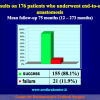
Results: The average follow-up was 53 mo. Of 375 cases, 313 (83.5%) were successful and 62 (16.5%) failures. Of 165 ARs, 150 (90.9%) were successful and 15 (9.1%) failures. Of 40 AARs, 24 (60%) were successful and 16 (40%) failures. Of 170 OGTs, 139 (81.8%) were successful and 31 (18.2%) failures. The AR showed statistically significant higher success rate compared to OGT (p=0.023) and AAR (p=0.0001). Of 47 PSGs, 28 (59.6%) were successful and 19 (40.4%) failures. Of 163 OMGs, 135 (82.8%) were successful and 28 (17.2%) failures. This difference was statistically significant (p=0.002).
Conclusions: One-stage bulbar urethroplasties showed an overall 83.5% success rate. The AR showed the higher success rate compared to the OGT or AAR. OMGs (82.8% success rate) perform statistically better than PSGs (59.6% success rate).

Article n° 13
Barbagli G, De Stefani S, Annino F, De Carne C, Bianchi G.,
Muscle- and Nerve-sparing Bulbar Urethroplasty: A New Technique.
Eur Urol 2008; 54:335 – 343

Background: To describe a new surgical technique for the repair of bulbar urethral strictures to preserve the bulbospongiosum muscle and its perineal innervation.
Objective: Surgical steps of muscle- and nerve-sparing bulbar urethroplasty are described. The outcome is provided regarding semen sequestration and postvoiding dribbling.
Design, Setting, and Participants: We performed the procedure in 12 patients (average age: 43.58 yr) with bulbar urethral strictures (average stricture length: 4.47cm).
Surgical Procedure: Six patients underwent urethroplasty using a ventral oral mucosal onlay graft, and six patients underwent urethroplasty using a dorsal oral mucosal onlay graft. In all patients, the surgical approach to the bulbar urethra was made avoiding dissection of the bulbospongiosum muscle from the corpus spongiosum and leaving the central tendon of the perineum intact.
Measurements: Clinical outcome was considered a failure when any postoperative instrumentation was needed. The primary outcome examined the technical feasibility of the muscle- and nerve-sparing bulbar urethroplasty. The secondary outcome examined the presence or absence of postoperative postvoid dribbling and semen sequestration using a nonvalidated questionnaire (Appendix).
Results and Limitations: In all patients, postoperative voiding cystourethrography was performed 3 wk after surgery and no urethral sacculation was evident. Urethrography were repeated after 6 mo and 12 mo. No postvoid dribbling or semen sequestration was demonstrated in all patients at 6 mo and 12 mo after surgery. No patient showed stricture recurrence. The average follow-up was 15.25 mo (range 12 mo to 26 mo, median 13.5 mo).
Conclusions: Bulbar urethroplasty preserving the bulbospongiosum muscle, the central tendon of the perineum, and the perineal nerves is a safe, feasible, minimally invasive alternative to traditional bulbar urethroplasty.
1. Question: What kind of anesthesia is used for this surgery?
Answer: General anesthesia with nasal intubation.


2. Question: How many hours does the surgery take?
Answer: About 2 hours.


3. Question: Are there any risks concerning erection, fertility and urinary incontinence after the surgery?
Answer: No, there aren’t.


4. Question: How many days of hospital recovery are expected following the surgery?
Answer: In general, from 5 to 7 days.


5. Question: How long will I have to use a catheter after the surgery?
Answer: The urethral catheter must stay in place for four weeks after the surgery, when the first post-operative voiding urethrography is done.


6. Question: Are there any particular recommendations during convalescence?
Answer: During convalescence, the use antibiotics until the catheter is removed is suggested. Avoiding long car trips, as well as heavy labor and sexual and sports activities are also suggested.


7. Question: When will I be able to resume my working, sexual and sports activities?
Answer: All these activities can be gradually resumed about 30 days after the removal of the catheter.


8. Question: Can I ride a bike or a motorcycle immediately after the surgery?
Answer: It is not recommended to ride bikes, motorcycles or horses after the surgery.


9. Question: What kinds of foods and drinks should be avoided after the surgery?
Answer: Beer and sparkling wines should be avoided, as well as large quantities of chocolate, cocoa, nuts and shellfish.